5. Colour

Photoreceptor neurons, whether rods or cones, either release glutamate or they don’t, there’s no middle ground. They are not equally responsive to light of different wavelengths, but the wavelength isn’t captured by the output, it’s just a yes/no kind of deal. Collectively, the pattern of releases from such a neuron tells you whether it is being stimulated more or less strongly, but it doesn’t tell you if the stimulation is strong because it’s at a wavelength the neuron is more sensitive to or just because there’s a lot of light — the releases would look the same either way. To be able to separate those cases, we would need more information. Fortunately, at least for most of us, more information is available.

The spectral responses of the different kinds of photoreceptors are illustrated below. (Note that each curve’s vertical extent is normalised so the peak sensitivity is at 100% in each case — rods are still way more sensitive that cones, but we don’t need to care about that right now.)

Based on Wikimedia Commons image by user Francois~frwiki, licensed CC BY-SA 4.0

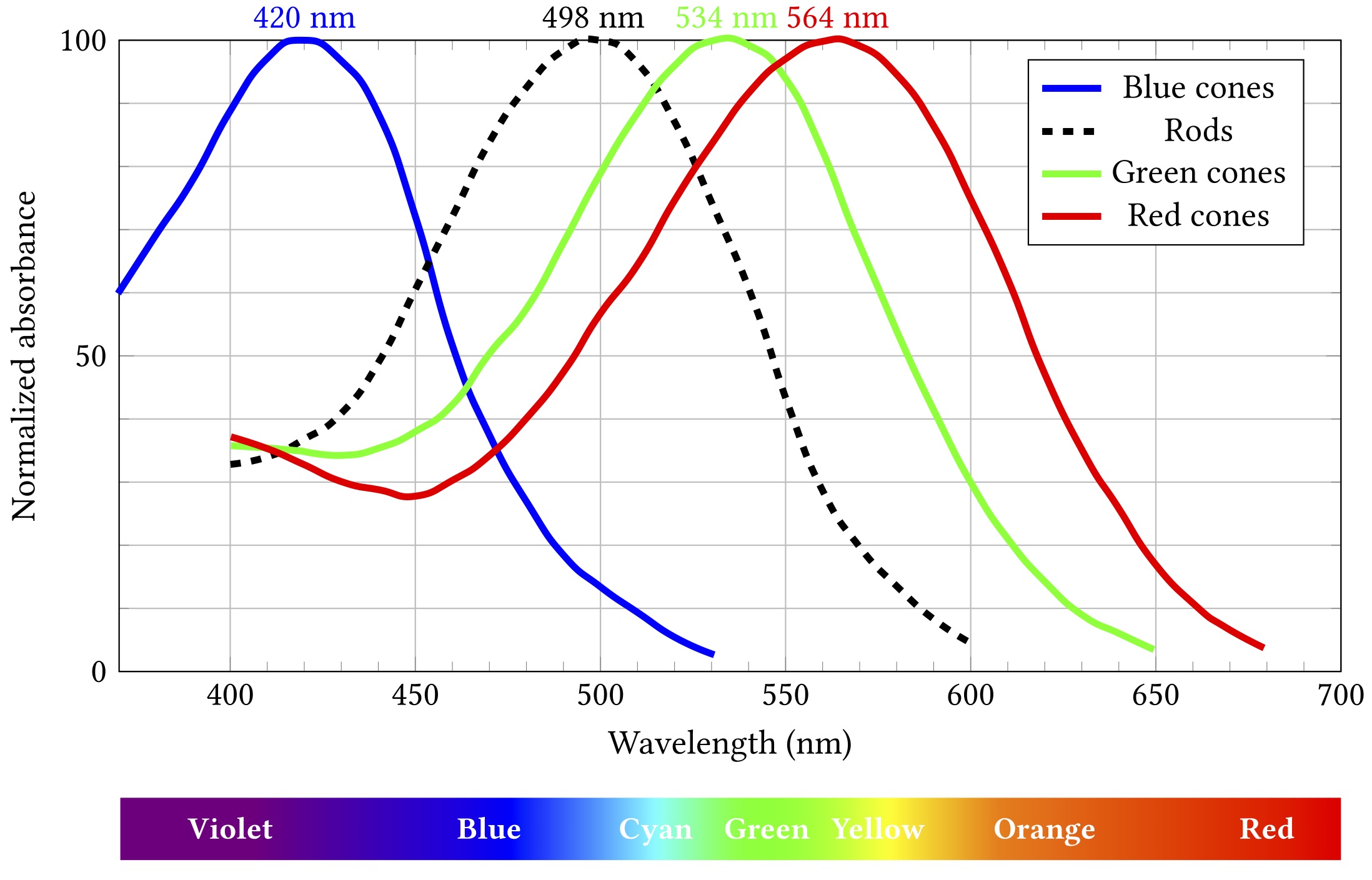
Importantly, while all rods have the same sensitivity, cones come in three different flavours with different absorption spectra. These flavours are sometimes termed blue, green and red, as in the diagram, but are also more accurately, if less picturesquely, known as short, medium and long. If cones of all three types are illuminated by light of the same colour, they will exhibit differing responses — and the differences can be used spectroscopically to determine what colour the light was.

At least, kinda.

Let’s say some blueish light of about 480 nm comes along:

Stimulation by 480 nm blue light

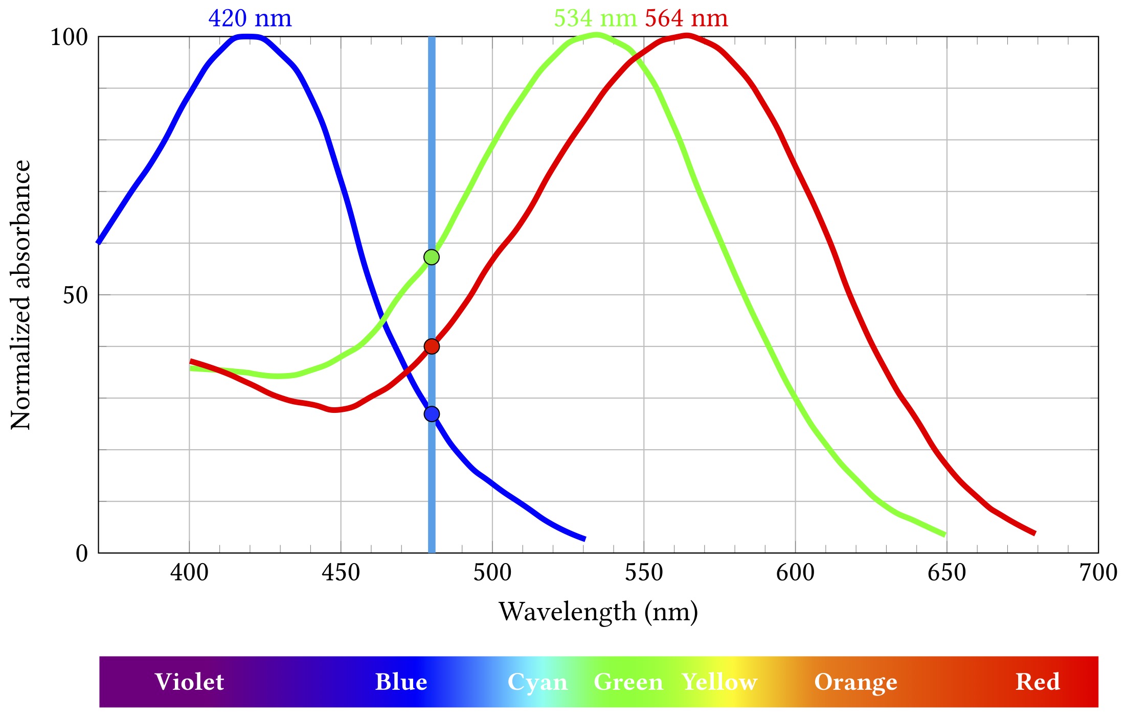
The short wavelength “blue” cones are actually stimulated least by this light, the long “red” cones a bit more, and the medium “green” ones most of all. Importantly, this particular combination of relative stimulation levels does not occur for any other wavelength, and so we can infer that the incoming light is that blueish 480 nm.

Except.

I mean come on, you’re reading this on an RGB screen, how much surprise can I honestly expect to milk out of it?

That uniqueness of inference only works if we know the light is all the same wavelength. That is almost never true, but it’s not a horribly misleading assumption when what you’re seeing started off as broad spectrum white light from the sun and then bounced off a bunch of objects that absorbed a lot of those wavelengths and just left some narrow band to hit your eyes. Which would mostly have been the case most of the time for most of the history of life on Earth. So it’s a pretty decent model for evolution to have hardwired into our biology.

Obviously these days we spend rather a lot of time looking at light from artificial sources for which the model doesn’t hold at all. But — and this is really pretty fortunate for a big chunk of modern human culture and technology — our visual system is still stuck on the veldt, looking for lions or whatever. We can bamboozle it with cheap parlour tricks — and we have gotten really good at those tricks.

You all know how this works: take three different light sources that mostly stimulate the long, medium and short cones respectively. Call ’em, oh I don’t know, red, green and blue? Shine them in the right proportions on your eyes and behold: suddenly you’re seeing orange or turquoise or mauve. Those wavelengths aren’t actually there — does mauve even have a wavelength? — but that doesn’t make your perception of them any less real.

We call these ingredients primary colours, but in what sense are they primary? There’s nothing special about red, green and blue from the point of view of light. Wavelengths don’t mix to make other wavelengths. Violet light is violet, it’s not some mixture of blue and red. Except: what is violetness anyway? As noted back in part 2, colour isn’t a property of light, it’s a perceptual property. I can confidently assert that 425 nm light isn’t a mixture of red and blue, but violet is whatever I see it is.

So primary colours really are primary, in the sense that we can use them to stimulate the perception of (very roughly speaking) any other colour, including some that don’t really exist in the visible spectrum at all. Their primariness — like colour itself — is a product of our physiology and neural processing.

Sing out, Louise. You’ve heard this song before, you probably know the words by now.

And you also know this one: the mechanics of visual perception are complex and fragile and things can go awry.

Colour perception is not universal and you cannot rely on everyone perceiving colours the same way you do — whatever way that is. Various mutations affect the cone cells, leading to a variety of colour vision deficiencies — from somewhat attenuated ability to distinguish between some colours to (rarely) complete monochromacy. These mutations are often sex-linked — ie, the affected proteins are on the X chromosome — so they run in families and mostly affect males.

This can be a problem because colour is a really handy and widely used design feature and popular carrier of meaning. Red means stop, green means go, yellow means go really fast. If you’re constructing an interface or visualising data, you’re probably going to want to put colour to work. Just consider the graphs above — colour is the key distinguishing feature of the three cone lines. So it’s a teensy bit problematic that up to 8% of the men reading this (pretend for a moment than anyone is going to read this) might struggle to tell two of them apart.

(Okay, yes, I’m wrapping this chapter with a rhetorical flourish and it’s really a lie — those particular lines are distinguishable by luminance as well as colour, so 8% of men will probably muddle through — but the point stands more broadly, just go with it.)

4. Retina

The retina is the thin tissue layer on the inside rear of the eyeball that is responsible for transduction of incoming light into electrochemical neural signals and also for several additional integration and processing steps on those signals. It is emphatically not just a simple sensor and indeed is considered to be part of the central, rather than peripheral, nervous system — an extension of the brain.

The retina is organised depthwise into several functional layers containing different types of neurons. These layers are inverted in humans and other vertebrates, meaning that processing proceeds from back to front. Light passes through all the layers, is then detected, and then the transduced nerve signals propagate back up through the layers, getting combined and filtered along the way. The results of all that processing then get delivered to the brain, but note that at that point we’re all the way to the front of the retina — the opposite side from the brain — so the top layer axons — the output cables that go to make up the optic nerve — need to pass back through the retina, making a hole with no detectors — the blind spot.

There are two different types of photoreceptive neurons in the back layer, known as rods and cones after the shapes of their outer segments (the bit of the cell where the actual photoreceptors live). These differ in their spatial distribution and connectivity and in their sensitivity to light — we’ll come back to this shortly.

Unusually, the rods and cones are depolarised at rest — that is, in the absence of a light stimulus the voltage difference across their membrane is relatively low, causing the neuron to release its neurotransmitter — glutamate — frequently. Incoming photons interact with photosensitive proteins in the outer segment, inducing a conformational change that in turn causes a cascade of signalling events that lead to hyperpolarisation of the neuron, making the interior more negative, which reduces its glutamate release. The changing release rate is received and processed by the next layer of neurons, the bipolar and horizontal neurons, and passed on in turn to the uppermost layer of retinal ganglion and amacrine neurons, which aggregate and process the visual information further before sending the resulting signals onward to the brain.

The rods and cones are distributed extremely non-uniformly over the retina. There’s a small area in the centre of the eye’s field of view known as the fovea, which contains only cones, no rods at all, and they’re packed in very densely. Outside the fovea there’s a mixture of both rods and cones, with the rods dominating almost everywhere — there are lot more rods than cones overall, of the order of 100 million compared to a mere 6 million or so cones. Both types become much sparser towards the periphery, and of course in the blind spot, which is about 20° nasal of the fovea, there are none.

In total there are about 100 times as many rods and cones as there are ganglion cells, so the signals going from the ganglions to the brain for visual processing can’t be just one-to-one reportage of the raw light detection. Rather, the data are summarised and transformed to pick out features of interest or smooth out noise or amplify weak signals. This funnelling of the photoreceptor signals into aggregated ganglion outputs is known as convergence — multiple photoreceptor neurons converge on each ganglion cell — and again it is not at all uniform across the eye. Rods in the periphery exhibit high convergence, with a single ganglion cell combining signals from hundreds or thousands of distinct receptors, improving sensitivity in those regions at a cost of spatial resolution. On the other hand, the densely packed cones of the fovea have very low convergence, preserving the fine spatial detail of detection and allowing for much greater visual acuity.

Rods are much more sensitive to light than cones. In bright conditions the cones respond strongly, allowing fine detail to be observed. But in dim conditions the fovea is pretty unresponsive, and vision is dominated by the high convergence periphery, meaning the acuity is much lower, and some objects may not be picked up by central vision, becoming only visible from the corner of your eye. Spooky!

Cones are also responsible for colour detection, as we’ll discuss next post, whereas rods do not distinguish colour — so night vision tends to be pretty monochromatic.

Exposure to light leads to desensitisation of the photoreceptors because the photosensitive pigments that do the detection are bleached in the process. These are continually replenished, but the process takes time. So the overall sensitivity of the retina is not fixed, it depends on how much light has been seen lately. If you’ve been out in bright daylight and go into a dark room, your sensitivity will have been depleted by the brightness and you won’t be able to see well. Whereas if you’ve been in the dark a long time, your receptor population will be fully replenished and you’ll have maximum sensitivity. This replenishment is known as adaptation — the eye has adapted to the darkness and is better able to see. Subsequent exposure to a lot of light will once again bleach the receptor pigments and reduce sensitivity. The time course of adaptation comes from the interplay between the rate of bleaching and the rate of replenishment.

As well as just aggregating the incoming signals, ganglion cells also pick out local spatial features, in particular brightness differences — edges — within the patch of retina whose photoreceptor cells that converge onto them. This patch is known as the receptive field — the region of the incoming image to which the neuron is receptive. (This concept is not limited to ganglion cells and we’ll encounter it throughout the processes of visual perception.)

Most retinal ganglion cells exhibit a centre-surround organisation to their receptive fields, whereby the response of the ganglion cell depends on the difference in activity at the centre of the receptive field and that in the surrounding region. The mechanism for this is lateral inhibition — the signal evoked by one region of photoreceptors is inhibited by the the signals from its neighbours. Ganglion cells may be off-centre — responding maximally to a dark middle and bright surround — or on-centre — responding to a bright centre and dark surround.

This kind of feature extraction isn’t a physiological necessity — the neurons could be wired up in arbitrary other ways. It’s a matter of perceptual utility. Edges are potentially important markers of stuff happening in the world — where one object begins and another ends. They represent things we might want to interact with, or run away from. Regions of uniform illumination evoke less response, because in some sense they are less perceptually interesting.

Once again, we note that perception is an active, cognitive process, rather than a passive consumptive one. Even before we leave the eye we’re already sifting and selecting and making value judgements.

3. Eyes

A basic camera consists of an aperture to control the amount of light getting in, usually adjustable by means of a diaphragm, a lens or system of lenses to capture and focus the light, and some form of chemical or electronic detector — all wrapped up in a dark box or chamber — from which the term camera comes — to stop light getting to the detector other than through the aperture & lens. All of these elements are also present in the eyes of many animals, including us.

The eye’s aperture is the pupil, adjusted by the sphincter and dilatory muscles of the iris to let in more light in dim conditions, less when it is bright.

The eye has two lens elements. The more rigid, fixed focal length cornea does the bulk of the focusing, while the softer, deformable lens provides adjustability. A ring of ciliary muscle around the lens can contract to increase the lens curvature, decreasing its focal length and allowing nearer objects to be in focus. This reshaping of the lens is known as accommodation.

The distances that an individual can bring into focus range from the far point, when the ciliary muscles are fully relaxed, to the near point when they are maximally contracted. These distances depend on the shape of the eye, the focusing power of the cornea and lens, the deformability of the lens and the strength of the ciliary muscles.

Impairments of focal range are very common. People with myopia or nearsightedness can’t focus on distant objects — their far point is significantly nearer than infinity and light from beyond that point comes to focus in front of the retina. Those with hyperopia or farsightedness can’t focus on nearby objects, the light from which comes to focus behind the retina. Both of these conditions can be corrected with glasses or contact lenses, introducing extra focusing elements in the optical path to change the baseline focusing power of the whole system. As people age, their lenses become increasingly stiff and their ciliary muscles weaken, reducing their capacity to accommodate, shifting the near point further away. This age-related farsightedness is known as presbyopia, literally “old eye”, and it’s why so many people need reading glasses as they get older.

Eyes are directional and there are several other sets of muscles connecting the eyeball to its socket that enable eye movements of various kinds — importantly, these movements are generally coordinated for both eyes. The types of movement include:

  • saccade — a fast skip or jump of the gaze from one static location (fixation point) to another, as when glancing between the faces of people you are talking to, or scanning along the words in a line of text
  • smooth pursuit — a steady continuous gaze shift that follows some moving object
  • vergence — movements that adjust the angle between the two eyes so that they are both pointing at the same target, rotating the angle closer together for nearby objects and further apart for more distant ones
  • gaze stabilisation — eye movements that compensate for changes in head position or view content to maintain a consistent field of view. A key example is the vestibulo-ocular reflex, which rotates and shifts eye position to oppose rotations and shifts of the head, driven by positional information detected in the inner ear.

Notably, most of these movements are not under conscious control. You can choose to look at some location, but you can’t choose to point your eyes in arbitrary directions independent of one another, or rotate your eyes about their view direction, or contract your irises. You can make your gaze trace a path in a series of saccades, but for smooth pursuit you pretty much need to watch something that’s actually in motion and let your brain move your eyes automatically. You can — just about — voluntarily shift focus or vergence, but it’s much much easier when you have some actual object to focus on, or at least can clearly imagine one.

Already we can see that the brain is imposing a huge amount of structure on the processes of vision that we just take for granted, and these percolate all the way through our visual perception. Things like saccades and the vestibulo-ocular reflex define the subjective experience of seeing — chopping up and smoothing out the world like a slick Hollywood production team, not some rough Blair Witch shakycam. And this is still just at the stage of the mechanics of light capture. We haven’t even got to the point of transduction yet. To do that, we need to talk about the eye’s detector component: the retina.

2. Optics & Images

You may recall from high school physics that light exhibits characteristics of both particles and waves. Different light attributes may be better thought of in terms of one or the other. For example, brightness is a function of the number of light particles — photons — arriving per unit time, whereas colour is a wave-like phenomenon, depending on the wavelength of those photons. For a given colour, increased brightness means having more photons arriving, not that the individual photons are themselves brighter.

(Note that I am being incredibly slipshod with the terminology here. As we’ll discuss in a little while, colour and brightness are not really attributes of the light at all, they are perceptual properties. But they’re a useful shorthand precisely because of that subjective familiarity.)

The energy of an individual photon depends on its wavelength or (reciprocally) frequency. Short wavelength photons have high frequencies and high energies, longer wavelengths have lower frequencies and lower energies. High energy photons can be damaging to biological tissues; lower energies can be harder to detect and broadly require larger sensory apparatus to distinguish from noise. That roughly 400 to 700 nm wavelength range of visible light represents a sort of Goldilocks zone of relatively harmless detectability in the context of human physiology. Visibility is not some physical property of light, it’s just how things are for us. Other animals will have different sensitivity ranges.

Light travels in straight lines at — famously — a constant speed. In a vacuum, that is. Matter complicates the picture in various ways. Usefully, we can think of the speed varying according to the medium through which the light is passing. Changes in medium induce speed changes and these lead to path changes. We can see this in effects such as heat haze, where fluctuating air density gives rise to wobbling. But in particular, crossing over distinct boundaries between different media — say between the air and a piece of glass — can alter the direction of the light, which we call refraction. This turns out to be super important.

If we want to use light as a medium for gathering information about the outside world, it’s helpful to know where it’s coming from. It’s not absolutely essential — there may still be some value in getting a general impression of aggregate lightness or darkness, and there are animals that do just that, but that only really scratches the surface of what we tend to think of as “vision”.

Assuming we have some kind of light detector — maybe some silver nitrate on a film or plate, a photodiode, or a nerve cell containing photosensitive pigments, whatever — if light reaches it simultaneously from different locations in the world, the information from each location will just pile up together in the detector’s response as an indistinguishable blur and we won’t be able to unpick which bit relates to what. What we would like is some form of spatial organisation, so that the light from one place all goes to the same detection point, while light from other places goes to other points.

One way to do this is to restrict the possible straight line paths of the light by forcing it all through the same point in space — a pinhole aperture. All the light arriving at any detector must be coming from exactly the direction of the pinhole because there’s nowhere else to go. This kind of setup is nice and simple and does occur in nature, especially in simpler organisms, requiring a lot less physiological machinery to evolve and build. But it has the disadvantage of capturing only a tiny fraction of the available light, discarding a lot of potentially useful information and reducing signal to noise.

An alternative is to have a larger aperture and use refraction through the curved surface boundaries of a lens to differentially bend the light passing through it, bringing the light to focus at the detector. Lens systems are harder to build than pinholes, both for human engineers and for biology, but they allow the capture of more light and more information.

The detector at a focused point has good information about one specific external position. If we have many detectors at many locations, that gives us a spatial map of optical information over a whole visual field — which is to say, an image.

Of course, focus is not perfect and not the same everywhere. Lenses will only bring to focus some parts of the external world — some range of distances from the lens. Detectors at well focused points will get strongly localised information, those where the focus is less good will receive light from a wider range of external regions and be correspondingly more imprecise, more blurry.

We are probably all familiar with this kind of locally focused imaging from using cameras, which combine a lens system for light gathering and focusing with some kind of spatial array of detectors — like a film frame or CCD — to perform the actual image capture.

Human vision is quite unlike photography in various important ways, some of which we’ll get into. But there are also enough structural similarities in the early stages that cameras are at least analogically useful as a model for starting to think about eyes.

The Vision Thing

[To bully myself into actually writing the lecture on visual perception I’m due to deliver in January, I’ve decided to try drafting the wretched thing as a series of blog posts. This is probably a very bad idea. It’s a two hour lecture, so there could potentially be quite a few of these posts, but also the chance of bailing is pretty high. Whatevs. Here goes nothin’.]

1. Preamble

To start with the bloomin’ obvious: vision is the process — the collection of processes — by which we gather information about the external world through the medium of light. To be precise, like Thomson and Thompson, that’s the medium of visible light — a relatively narrow band of electromagnetic radiation of wavelengths roughly 400 to 700 nm.

Light is a pretty great medium for gathering information — almost the definitive one. It propagates really fast over long distances, even through a vacuum. It travels, at least to a reasonable approximation, in straight lines. It interacts with matter and participates in chemical reactions, making it possible for biology to come up with mechanisms for detecting it. And it’s plentiful. There’s a lot of it about, at least during the day, thanks to an enormous nuclear furnace burning in the sky.

Vision is so useful that it has evolved numerous times over the history of life on Earth, with a fair bit of variation in the implementation details. There are a bunch of different structures and configurations of optical sense organs — the eyes of a fly or a flatworm or a scallop are quite different from our own — although some of the underlying biochemical components are pretty well conserved. We’ll touch on those in a little while.

Vision is also so useful that it’s a bit overpowering. It’s the dominant sense for most humans, one on which we rely heavily, one that radically shapes our understanding of the world, both literally and metaphorically.

The vocabulary of vision pervades our language, encompassing much more than mere optical detection: we say “oh, I see” meaning “I understand“, or “let’s see” meaning “we want to find out” or “we’ll see about that” meaning “I will act to prevent it.” The word vision doesn’t just refer to the sense of sight, but extends to intellectual brilliance, to clarity and drive, to divine revelation. We describe a great creator as a visionary, someone who predicts the future as a seer, or a clairvoyant — literally one who sees clearly.

Much of human culture revolves around the visual — painting and drawing, photography and movies, games. Practical necessities like food and clothing are wrapped up in visual aesthetics. The written word is primarily mediated — read — through our eyes. We have all manner of advanced technology dedicated to the enhancement of vision — microscopes and cameras and telescopes — and to the production of visual stimuli — from paper and pencils to printing and screens to HUDs and holograms and VR headsets. It is one of the major ways we interact with computers and software, often the main component of a user interface.

At the same time, vision is complex and fragile and lots of things can go wrong with it. Visual impairments are extremely common. People may have difficulty with focus or resolution close up or far away or — especially when you get to be ancient like me — both. People — particularly men — may be unable to distinguish some or all variations in colour. People may be unable to see in dim or bright conditions. They may lose parts or all of their visual field to obstructions in the eyes or degeneration of the sensory tissues or loss or damage to perceptual pathways. Some of these problems can be readily mitigated with technology — very many of us wear glasses or contact lenses, whether all the time or for specific tasks — other problems not so much.

So, given both vision’s centrality to human endeavours and its frequent failings, it is important to understand how it works and how it goes wrong, and try to find ways we can make the most of it while also maximising accessibility.